| 产品编号 | bsm-33033M |
| 英文名称 | GAPDH-Loading Control |
| 中文名称 | 3-磷酸甘油醛脱氢酶(内参)单克隆抗体 |
| 别 名 | 38 kDa BFA-dependent ADP-ribosylation substrate; Aging-associated gene 9 protein; BARS-38; cb609; EC 1.2.1.12; G3PD; G3PDH; GAPD; Glyceraldehyde 3 phosphate dehydrogenase;Glyceraldehyde 3 phosphate dehydrogenase liver;Glyceraldehyde 3 phosphate dehydrogenase muscle; KNC-NDS6; MGC102544; MGC102546; MGC103190; MGC103191; MGC105239; MGC127711; MGC88685; OCAS, p38 component; OCT1 coactivator in S phase, 38-KD component; wu:fb33a10. |
| 研究领域 | 肿瘤 细胞生物 免疫学 神经生物学 新陈代谢 |
| 抗体来源 | Mouse |
| 克隆类型 | Monoclonal |
| 克 隆 号 | 4F8 |
| 交叉反应 | Human, Mouse, Rat, Chicken, Dog, Pig, Rabbit, Sheep, Hamster, Monkey, |
| 产品应用 |
WB=1:5000-20000 IHC-P=1:100-500 ICC=1:100 (石蜡切片需做抗原修复) not yet tested in other applications. optimal dilutions/concentrations should be determined by the end user. |
| 分 子 量 | 38kDa |
| 细胞定位 | 细胞核 细胞浆 细胞膜 |
| 性 状 | Liquid |
| 浓 度 | 1mg/ml |
| 免 疫 原 | Recombinded Human GAPDH : |
| 亚 型 | IgG |
| 纯化方法 | affinity purified by Protein G |
| 储 存 液 | 0.01M TBS(pH7.4) with 1% BSA, 0.03% Proclin300 and 50% Glycerol. |
| 保存条件 | Shipped at 4℃. Store at -20 °C for one year. Avoid repeated freeze/thaw cycles. |
| PubMed | PubMed |
| 产品介绍 |
Loading Control Function: Has both glyceraldehyde-3-phosphate dehydrogenase and nitrosylase activities, thereby playing a role in glycolysis and nuclear functions, respectively. Participates in nuclear events including transcription, RNA transport, DNA replication and apoptosis. Nuclear functions are probably due to the nitrosylase activity that mediates cysteine S-nitrosylation of nuclear target proteins such as SIRT1, HDAC2 and PRKDC. Glyceraldehyde-3-phosphate dehydrogenase is a key enzyme in glycolysis that catalyzes the first step of the pathway by converting D-glyceraldehyde 3-phosphate (G3P) into 3-phospho-D-glyceroyl phosphate. Subunit: Homotetramer. Interacts with TPPP; the interaction is direct. Interacts (when S-nitrosylated) with SIAH1; leading to nuclear translocation. Interacts with RILPL1/GOSPEL, leading to prevent the interaction between GAPDH and SIAH1 and prevent nuclear translocation. Interacts with EIF1AD, USP25, PRKCI and WARS. Subcellular Location: Cytoplasm, cytosol. Nucleus. Cytoplasm, perinuclear region. Membrane. Note=Translocates to the nucleus following S-nitrosylation and interaction with SIAH1, which contains a nuclear localization signal. Postnuclear and Perinuclear regions. Post-translational modifications: S-nitrosylation of Cys-152 leads to interaction with SIAH1, followed by translocation to the nucleus. ISGylated (Probable). Sulfhydration at Cys-152 increases catalytic activity. Similarity: Belongs to the glyceraldehyde-3-phosphate dehydrogenase family. SWISS: P04406 Gene ID: 2597 Database links: Entrez Gene: 374193 Chicken Entrez Gene: 2597 Human Entrez Gene: 100042025 Mouse Entrez Gene: 14433 Mouse Entrez Gene: 317743 Zebrafish Omim: 138400 Human SwissProt: P00356 Chicken SwissProt: P04406 Human SwissProt: P16858 Mouse SwissProt: Q5XJ10 Zebrafish Important Note: This product as supplied is intended for research use only, not for use in human, therapeutic or diagnostic applications. GAPDH蛋白几乎在所有组织中都高水平表达,广泛用作Western blot蛋白质标准化的内参,是很好的内参抗体。 GAPDH 作为管家基因在同种细胞或者组织中的蛋白质表达量一般是恒定的。在实验中,可能存在总蛋白浓度测定不准确;或者蛋白质样品在电泳前上样时产生的样品间的操作误差;这些误差需要通过测定每个样品中实际转到膜上的GAPDH的含量来进行校正,所以一般的western实验都需要进行内参设置。具体校正的方法就是将每个样品测得的目的蛋白含量与本样品的GAPDH含量相除,得到每个样品目的蛋白的相对含量。然后才进行样品与样品之间的比较。 甘油醛-3-磷酸脱氢酶(Glyceraldehyde 3 phosphate dehydrogenase,GAPDH)是糖酵解(glycolysis)过程中的关键酶。除了在胞质中作为糖酵解的酶以外,有证据表明哺乳动物细胞中的GAPDH参与了多种胞内生化过程,包括膜融合(membrane fusion)、微管成束(microtubule bundling)、磷酸转移酶(phosphotransferase)激活、核内RNA出核、DNA复制与DNA修复。一些生理因素,诸如低氧(hypoxia)和尿糖(diabetes),可以增加GAPDH在特定细胞中的表达。GAPDH存在于几乎所有的组织中,以高水平持续表达。 GAPDH(甘油醛-3-磷酸脱氢酶)是参与糖酵解的一种关键酶,由4个30-40kDa的亚基组成. |
| 产品图片 |
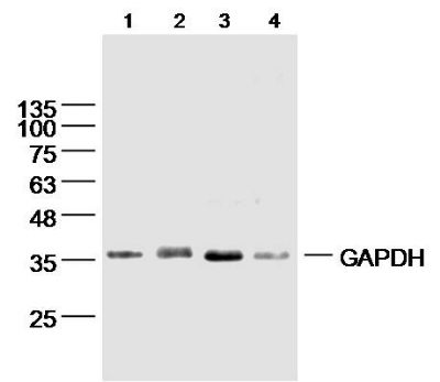
Sample:
Lane1: Skin (Mouse) Lysate at 40 ug Lane2: Testis (Mouse) Lysate at 40 ug Lane3: Adrenal gland (Mouse) Lysate at 40 ug Lane4: Lung (Rat) Lysate at 30 ug Primary: Anti-GAPDH (bsm-33033M) at 1/1 000 dilution Secondary: IRDye800CW Goat Anti-Mouse IgG at 1/20000 dilution Predicted band size: 38 kD Observed band size: 38 kD
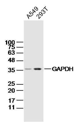
Sample:
A549 Cell Lysate at 25 ug 293T Cell Lysate at 40 ug Primary: Anti-GAPDH(bsm-33033M)at 1/5000 dilution Secondary: IRDye800CW Goat Anti-RabbitIgG at 1/20000 dilution Predicted band size: 38kD Observed band size: 38kD
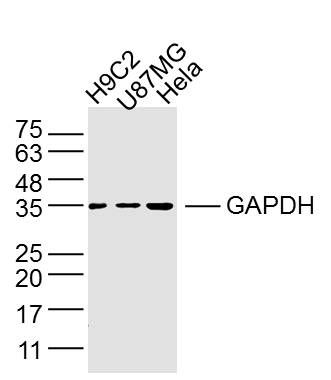
Sample:
H9C2 Cell (Rat) Lysate at 40 ug U87MG Cell (Human) Lysate at 40 ug Hela Cell (Human) Lysate at 40 ug Primary: Anti- GAPDH (bsm-33033M) at 1/2 000 dilution Secondary: IRDye800CW Goat Anti-Mouse IgG at 1/20000 dilution Predicted band size: 38 kD Observed band size: 35 kD
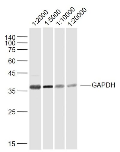
Sample:
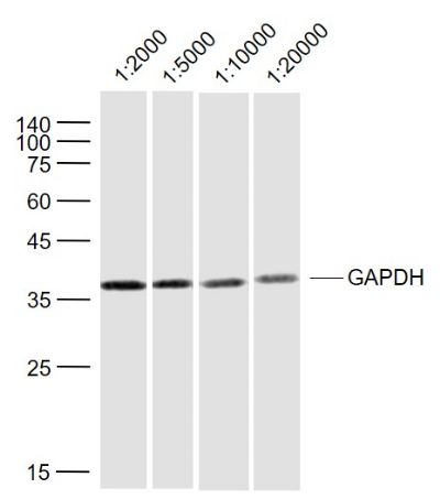
Lane 1: Cerebrum (Rat) Lysate at 40 ug Lane 2: Cerebrum (Rat) Lysate at 40 ug Lane 3: Cerebrum (Rat) Lysate at 40 ug Lane 4: Cerebrum (Rat) Lysate at 40 ug Primary: Lane 1: Anti-GAPDH (bsm-33033M) at 1/2000 dilution Lane 2: Anti-GAPDH (bsm-33033M) at 1/5000 dilution Lane 3: Anti-GAPDH (bsm-33033M) at 1/10000 dilution Lane 4: Anti-GAPDH (bsm-33033M) at 1/20000 dilution Secondary: IRDye800CW Goat Anti-Mouse IgG at 1/20000 dilution Predicted band size: 38 kD Observed band size: 36 kD
Sample:
Lane 1: Hela (Human) Lysate at 40 ug Lane 2: Hela (Human) Lysate at 40 ug Lane 3: Hela (Human) Lysate at 40 ug Lane 4: Hela (Human) Lysate at 40 ug Primary: Lane 1: Anti-GAPDH (bsm-33033M) at 1/2000 dilution Lane 2: Anti-GAPDH (bsm-33033M) at 1/5000 dilution Lane 3: Anti-GAPDH (bsm-33033M) at 1/10000 dilution Lane 4: Anti-GAPDH (bsm-33033M) at 1/20000 dilution Secondary: IRDye800CW Goat Anti-Mouse IgG at 1/20000 dilution Predicted band size: 38 kD Observed band size: 36 kD
Sample:
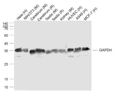
Lane 1: Hela (Human) Cell Lysate at 30 ug Lane 2: NIH/3T3 (Mouse) Cell Lysate at 30 ug Lane 3: Cerebrum (Mouse) Lysate at 40 ug Lane 4: Cerebrum (Rat) Lysate at 40 ug Lane 5: Testis (Mouse) Lysate at 40 ug Lane 6: Testis (Rat) Lysate at 40 ug Lane 7: Kidney (Mouse) Lysate at 40 ug Lane 8: HUVEC (Human) Cell Lysate at 30 ug Lane 9: A549 (Human) Cell Lysate at 30 ug Lane 10: MCF-7 (Human) Cell Lysate at 30 ug Primary: Anti-GAPDH (bsm-33033M) at 1/1000 dilution Secondary: IRDye800CW Goat Anti-Mouse IgG at 1/20000 dilution Predicted band size: 36 kD Observed band size: 36 kD
Sample:
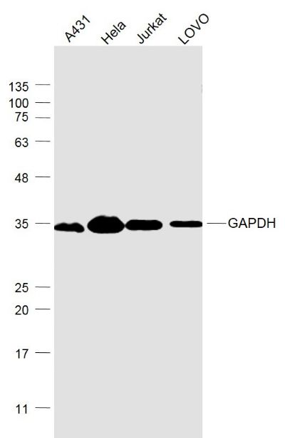
A431(Human) Cell Lysate at 30 ug Hela(Human) Cell Lysate at 30 ug Jurkat(Human) Cell Lysate at 30 ug LOVO(Human) Cell Lysate at 30 ug Primary: Anti- GAPDH (bsm-33033M) at 1/1000 dilution Secondary: IRDye800CW Goat Anti-Mouse IgG at 1/20000 dilution Predicted band size: 38 kD Observed band size: 35 kD
Sample:
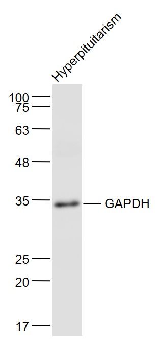
Hyperpituitarism (Mouse) Lysate at 40 ug Primary: Anti- GAPDH (bsm-33033M) at 1/5000 dilution Secondary: IRDye800CW Goat Anti-Mouse IgG at 1/20000 dilution Predicted band size: 38 kD Observed band size: 34 kD
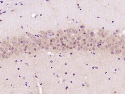
Paraformaldehyde-fixed, paraffin embedded (mouse brain); Antigen retrieval by boiling in sodium citrate buffer (pH6.0) for 15min; Block endogenous peroxidase by 3% hydrogen peroxide for 20 minutes; Blocking buffer (normal goat serum) at 37°C for 30min; Antibody incubation with (GAPDH-Loading Control) Monoclonal Antibody, Unconjugated (ascites of bsm-33033M-4E8) at 1:2000 overnight at 4°C, followed by operating according to SP Kit(Mouse) (sp-0024) instructions and DAB staining.
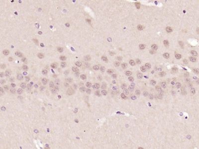
Paraformaldehyde-fixed, paraffin embedded (rat brain); Antigen retrieval by boiling in sodium citrate buffer (pH6.0) for 15min; Block endogenous peroxidase by 3% hydrogen peroxide for 20 minutes; Blocking buffer (normal goat serum) at 37°C for 30min; Antibody incubation with (GAPDH-Loading Control) Monoclonal Antibody, Unconjugated (ascites of bsm-33033M-4E8) at 1:2000 overnight at 4°C, followed by operating according to SP Kit(Mouse) (sp-0024) instructions and DAB staining.
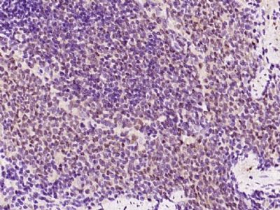
Paraformaldehyde-fixed, paraffin embedded (rat spleen); Antigen retrieval by boiling in sodium citrate buffer (pH6.0) for 15min; Block endogenous peroxidase by 3% hydrogen peroxide for 20 minutes; Blocking buffer (normal goat serum) at 37°C for 30min; Antibody incubation with (GAPDH-Loading Control) Monoclonal Antibody, Unconjugated (ascites of bsm-33033M-4E8) at 1:2000 overnight at 4°C, followed by operating according to SP Kit(Mouse) (sp-0024) instructions and DAB staining.
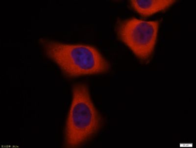
Tissue/cell:Hela cell; 4% Paraformaldehyde-fixed; Triton X-100 at room temperature for 20 min; Blocking buffer (normal goat serum, C-0005) at 37°C for 20 min; Antibody incubation with (GAPDH-Loading Control) monoclonal Antibody, Unconjugated (bsm-33033M) 1:100, 90 minutes at 37°C; followed by a CY3 conjugated Goat Anti-Mouse IgG antibody at 37°C for 90 minutes, DAPI (blue, C02-04002) was used to stain the cell nuclei.
|
1.《Multifunction Sr, Co and F co-doped microporous coating on titanium of antibacterial, angiogenic and osteogenic activities》
作者:Jianhong Zhou ,Lingzhou Zhao 影响因子:4.259
期刊:《Scientific Reports 6, Article number: 29069 (2016)》 PMID:27353337
作者:Jianhong Zhou ,Lingzhou Zhao 影响因子:4.259
期刊:《Scientific Reports 6, Article number: 29069 (2016)》 PMID:27353337
| 用户名: | (可为空) |
| E-mail: | *(必填) |
| 评价等级: |


|
| 评论内容: |
*(必填)
|
 中文
中文 
